Antibody-Drug Conjugate (ADC) Discovery and Development
Analytical tools for every phase of ADC development, from conjugation and internalization to Fc characterization, payload assessment, and bystander killing analysis.
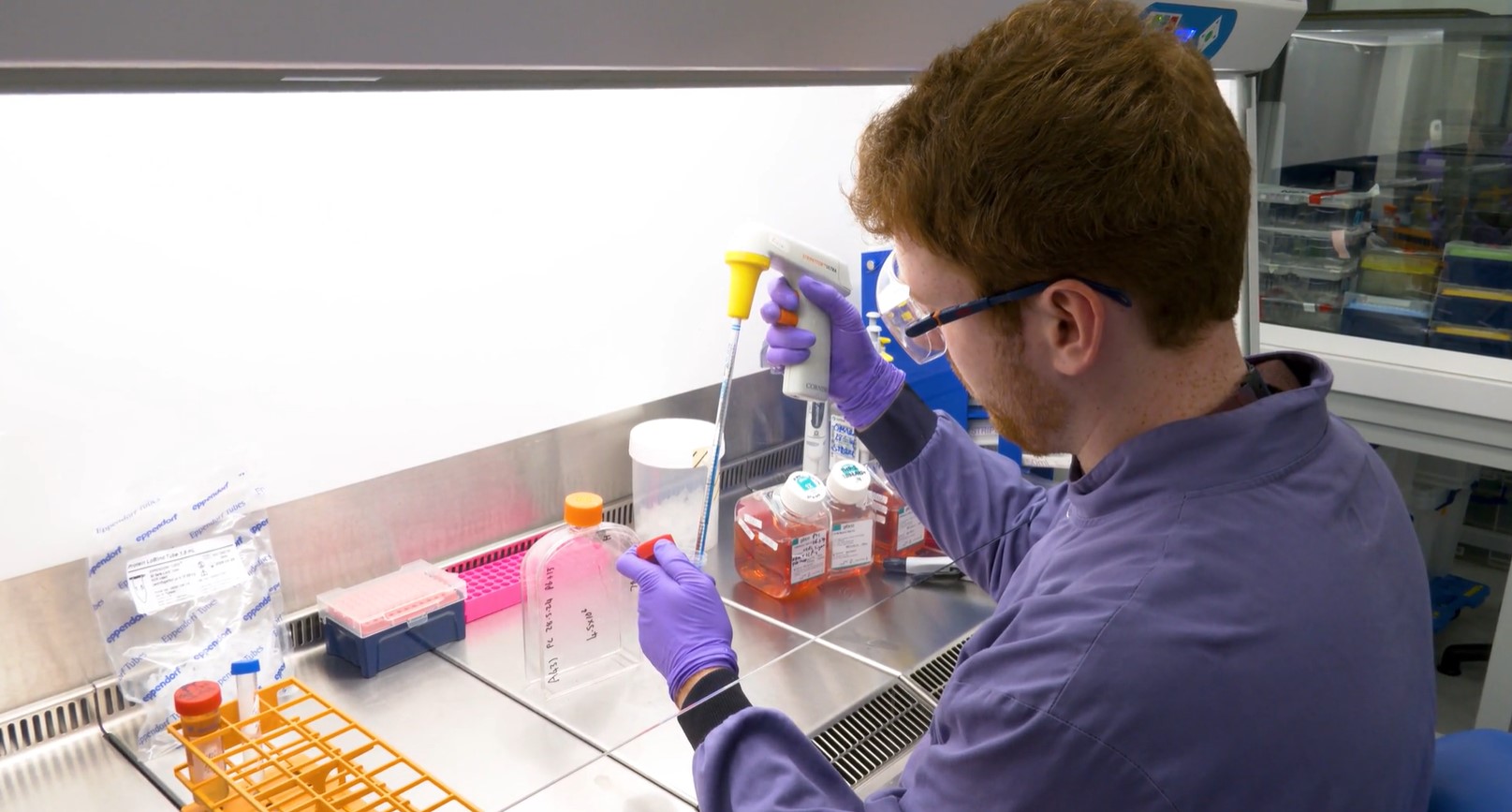
See how innovative assays like CellTiter-Glo® and RealTime-Glo™ streamline workflows, enhance insights, and drive advancements in ADC research.
Key Takeaways
- Promega provides validated bioluminescent assays across the ADC workflow: Antibody Engineering, Fc Effector Function, Internalization, Cytotoxicity, Payload Function, and Drug Release.
- Technologies including NanoLuc®, NanoBiT®, and HiBiT® Assays deliver superior signal-to-noise ratio and high-sensitivity readouts compared to fluorescent methods, enabled for real-time kinetic and endpoint readouts.
- Promega's Tailored R&D Solutions team offers custom assay development, screening, and mechanism-of-action studies to accelerate your ADC program.
What Is an Antibody-Drug Conjugate (ADC) and How Does It Work?
An antibody-drug conjugate is a targeted cancer therapeutic that links a monoclonal antibody to a cytotoxic payload through a chemical linker. The antibody selectively binds a tumor-specific antigen; once internalized, the linker releases the payload inside the cancer cell.
ADCs have three critical structural components: the antibody (engineered to bind a tumor-specific antigen), the linker (cleavable or non-cleavable, stable in circulation but designed to release upon internalization), and the payload (a potent cytotoxic molecule such as a microtubule inhibitor, DNA-damaging agent, or protein degrader).
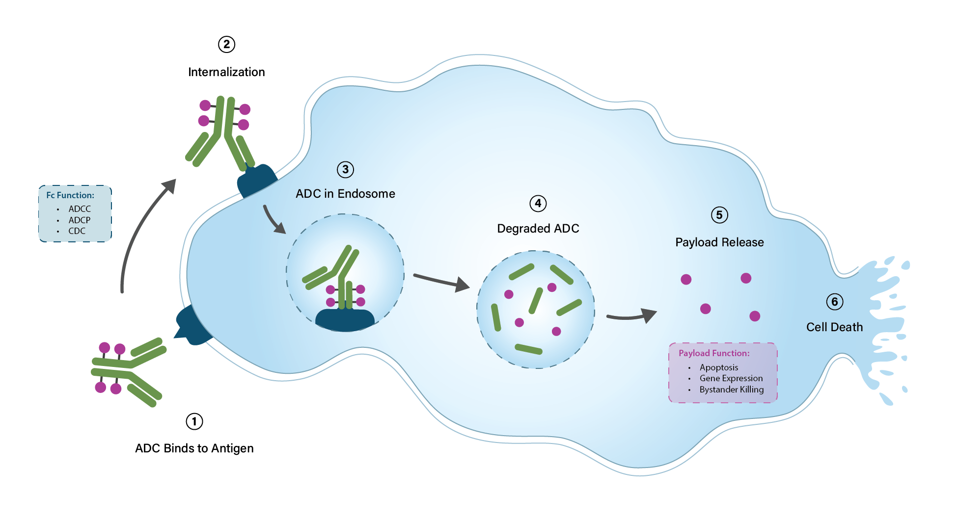
This modular architecture allows ADCs to deliver cell-killing potency with antibody-level targeting precision, achieving greater efficacy and reduced off-target toxicity compared to conventional chemotherapy. Beyond direct payload-mediated killing, ADCs can also engage Fc-mediated immune effector mechanisms including ADCC, ADCP, and CDC.
From Binding to Cell Death: An ADC Cellular Processing Pathway
Six steps, from antigen binding to cell death. Click a marked element for a preview. The resource opens only when you choose to.
Antibody Engineering
Before ADC internalization, several aspects of the antibody structure are engineered. Affinity, target antigen, Fc activity, and drug-to-antibody ratio, plus cleavable or non-cleavable linkers, influence downstream payload effects.
Download Resource »The Antibody Internalization Bioassay
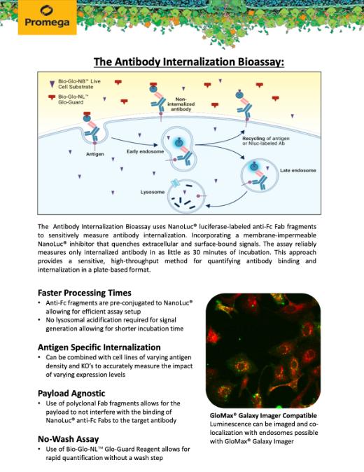
Antibody-drug conjugates (ADCs) combine the specificity of monoclonal antibodies with the potency of cytotoxic drugs to selectively kill cancer cells. To be effective, ADCs must be internalized and trafficked inside the cell to release their payload and avoid off-target effects.
ADC Degradation
Endosome acidification and subsequent lysosomal fusion lead to ADC degradation and payload activation. The payload is then free to exert its function by a variety of means, including DNA damage, protein interactions, or altering gene expression.
Read More »Direct, bystander and off-target ADC killing
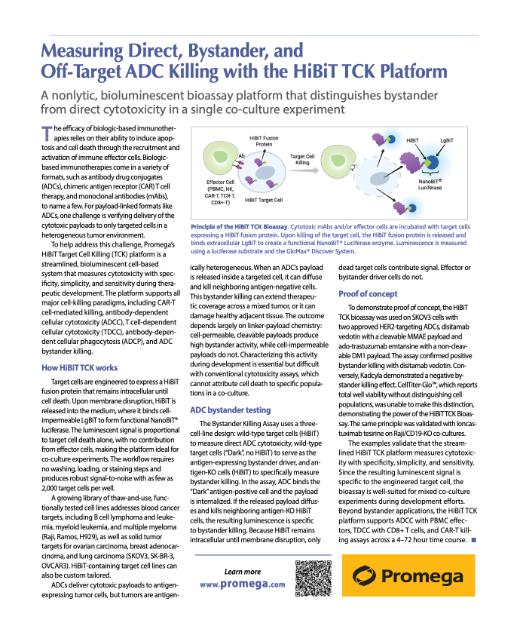
When an ADC's payload is released inside a targeted cell, it can diffuse and kill neighboring antigen-negative cells. This bystander killing can extend therapeutic coverage across a mixed tumor, or it can damage healthy adjacent tissue.
Cell Health Assays
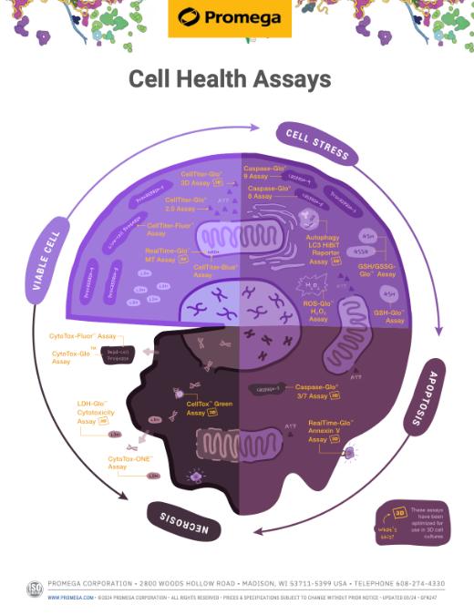
ADCs deliver cytotoxic payloads causing cell death, but understanding which pathway is critical. Promega cell health assays measure apoptosis, viability, and caspase activation to optimize ADC therapeutic windows.
Fc Effector Activity Bioassays
The ADC's Fc domain recruits immune effector functions: ADCC, ADCP, and CDC. A comprehensive workflow spanning binding characterization, functional readouts, and primary cell bridging enables developers to optimize Fc-region performance from lead generation through lot release.
Read More »New to ADC research? Our scientists can help you select the right assay workflow for your program.
How Do You Measure ADC Internalization?
Efficient internalization is a prerequisite for payload delivery. Promega offers two complementary approaches: real-time pH-reactive dye tracking and a pH-independent bioluminescent readout to quantify how effectively an ADC enters target cells.
pHAb Reactive Dyes: Real-Time Tracking
pHAb Reactive Dyes enable ADC researchers to precisely track antibody internalization into cells. These dyes fluoresce in acidic conditions, like those in endosomes and lysosomes, offering a quantitative measure of ADC delivery efficiency. This aids in evaluating and enhancing ADC therapeutic potential.
See more data in this publication: Homogeneous plate based antibody internalization assay using pH sensor fluorescent dye
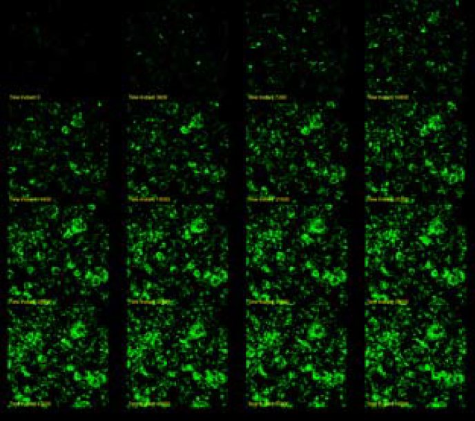
Trastuzumab (anti-HER2) was labeled with pHAb dye. SKBR3 cells were treated with 30nM Trastuzumab-pHAb dye. Image was captured every 60 minutes.
Antibody Internalization Bioassay
The Antibody Internalization Bioassay gives researchers an endpoint readout to monitor antibody internalization. The assay is based on an anti-Fc Fab labeled with NanoLuc® luciferase binding to a primary antibody or ADC therapeutic.
When the primary antibody binds to the target antigen, the complex is internalized, while unbound antibody is washed away. The detection reagent that includes a membrane-impermeable NanoLuc® inhibitor (Bio-Glo-NL™ Glo-Guard), a membrane-impermeable NanoLuc® inhibitor buffer (Bio-Glo-NL™ Glo-Guard Buffer, yellow shading in figure) and a membrane-permeable luciferase substrate is added to the cells. The resulting luminescent signal is primarily generated from the internalized Fab:primary antibody complex.
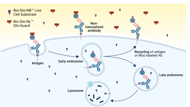
Accurately Compare Antibody Internalization Across Structural Variants in Adherent and Suspension Cells
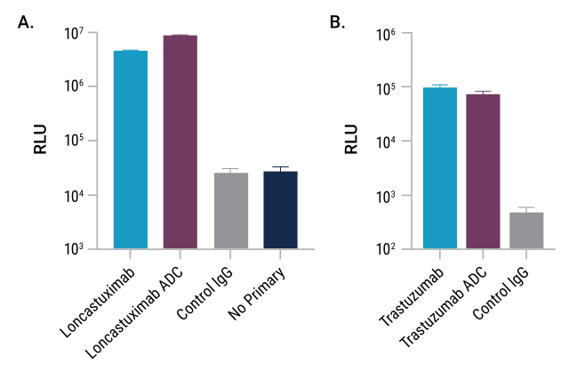
The Antibody Internalization Bioassay was used to quantify the internalization of two classes of ADCs and a non-conjugated version.
Panel A. Raji target cells were plated with CD19-specific antibodies loncastuximab, a loncastuximab ADC and a control IgG.
Panel B. SKOV3 target cells were plated with HER2-specific trastuzumab, a trastuzumab ADC and a control IgG. The Antibody Internalization Bioassay is amenable to measuring internalization in both suspension (Raji) and adherent (SKOV3) cell types.
How Do You Assess ADC Payload Function and Cytotoxicity?
Promega's multiplexable cell viability, cytotoxicity, and apoptosis assays provide real-time and endpoint readouts of how ADC payloads affect target cell survival and death mechanisms, providing critical data for optimizing therapeutic design.
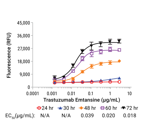
Cytotoxicity and Apoptosis Multiplexing
| Assay | What It Measures | Readout | Kinetics |
|---|---|---|---|
| CellTox Green™ Assay | Membrane integrity | Fluorescence | Real-time |
| CellTiter-Fluor™ Assay | Live cell protease activity | Fluorescence | Endpoint |
| Caspase-Glo® 3/7 | Caspase activation | Luminescence | Endpoint |
| RealTime-Glo™ Annexin V | Apoptosis & necrosis kinetics | Lum + Fluor | Real-time |
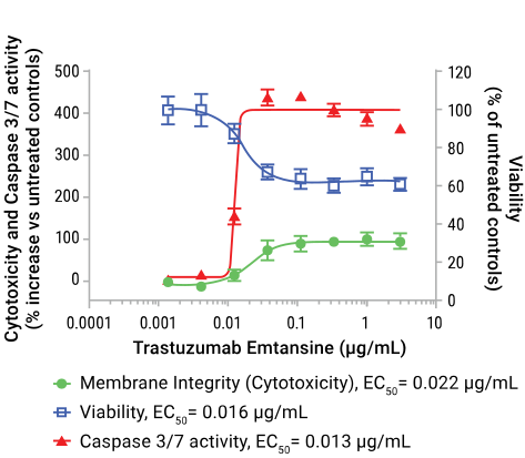
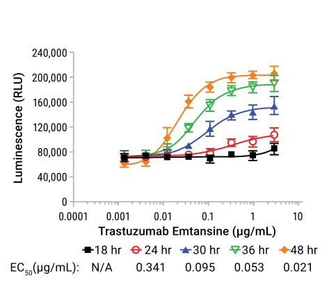
Immunogenic Cell Death (ICD)
Some ADC payloads induce immunogenic cell death, releasing DAMPs that activate antitumor immune responses. Lumit® Cytokine Immunoassays measure key pro-inflammatory cytokines (IL-1β, IL-6, TNF-α, IFN-γ) for evaluating the immune system's response to ADC therapies.
Next-Generation Payloads
ADC payloads are expanding beyond traditional cytotoxins. Explore tools for degrader (PROTAC) payloads and oligonucleotide/RNA-targeting payloads.
Developing a cytotoxicity assay for your ADC? Our team can help design the right multiplexed workflow.
How Does Promega Support ADC Engineering?
On-bead conjugation and purification with magnetic beads, combined with mass spectrometry-grade proteases, provide scalable workflows for ADC development and Critical Quality Attribute quantification.
On-bead Conjugation and Purification
Magne® Protein G and Magne® Protein A Beads are designed for the efficient and selective purification of antibodies. For ADC development, this technology facilitates the isolation of high-purity antibodies from complex biological mixtures and allows on-bead conjugation. The use of magnetic beads allows for a straightforward, scalable, and rapid conjugation and purification process. Our Field Support team, with expertise on HT platforms of all kinds, can program this chemistry for success in your lab.
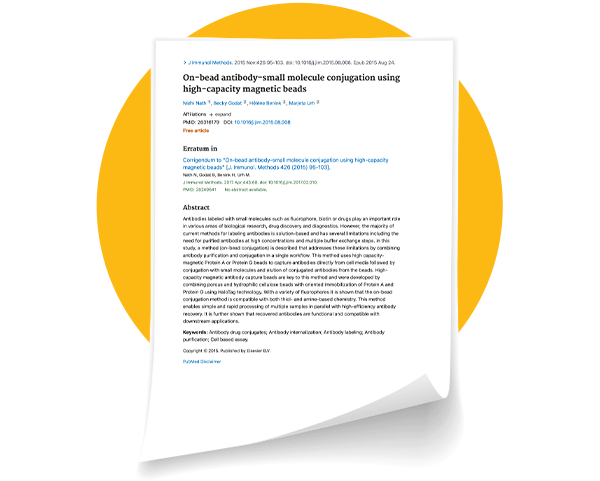
See data in this publication: On-bead antibody-small molecule conjugation using high-capacity magnetic beads.
Mass Spectrometry
Mass spectrometry (MS) is a critical tool for ADC research for its precision in characterizing, quantifying, and ensuring that Critical Quality Attributes (CQA) of these complex therapeutics are met. MS can be used to:
- confirm sequence of biotherapeutic protein, including ADC conjugation site
- reveal drug-to-antibody ratios
- accurately measure ADC concentrations in biological samples (preclinical and clinical)
- assess ADC stability and degradation
- ensure each ADC batch meets stringent quality standards and lot-to-lot consistency
- monitor host cell protein impurities
Learn more about our solutions for MS, including proteases and reference reagents.
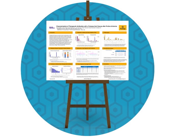
See data in this poster: Characterization of Therapeutic Antibodies with a ProAlanase
Proteases for ADC Characterization
Need help selecting the right protease for your ADC characterization workflow? Select the protease from the table below.
+++ Recommended ++ Conditional – Not recommended
| Enzyme | Cleavage Specificity | Conjugation Site Mapping | DAR Determination | Disulfide / Non-Reduced Mapping | Peptide Mapping (mAb Backbone) | Key ADC Advantage |
|---|---|---|---|---|---|---|
| Trypsin Platinum Recombinant MS-Grade | C-terminal of Arg and Lys (not before Pro) | Cleaner maps; recombinant purity eliminates chymotryptic noise at conjugation sites | Best-in-class for bottom-up DAR; zero non-specific activity | Alkaline pH risk; suppress scrambling with IAA | Zero non-specific activity; superior for RP-HPLC-UV and quantitative workflows | Recombinant design eliminates trace chymotrypsin that masks hydrophobic drug-containing peptides; upgrade for regulated ADC characterization and QC lot release |
| Trypsin/Lys-C Mix MS-Grade | C-terminal of Arg and Lys (sequential Lys-C then trypsin) | Improved digestion of hydrophobic ADC regions; Lys-C step resolves Lys-conjugated peptides more consistently | Preferred for bottom-up DAR; fewer missed cleavages means cleaner quantitation | Alkaline pH; use IAA to suppress scrambling | Fewer missed cleavages than trypsin alone; best overall mAb backbone sequence coverage | Sequential Lys-C + trypsin maximizes completeness in hydrophobic ADC regions where trypsin alone stalls; preferred single-enzyme upgrade from standard trypsin |
| IdeS / IdeZ IgG-Cleaving Enzymes | Single cut below IgG hinge (all human IgG1–4; IdeZ adds mouse IgG) | Domain-level only; cannot pinpoint specific residue | Generates F(ab')₂ + Fc with intact drug–linker; ideal for middle-up DAR; IdeZ adds mouse IgG2a/IgG3 | Non-reduced hinge region accessible but limited peptide coverage | Not for peptide mapping | Workhorse for middle-up ADC DAR: single, site-specific IgG cut under mild, native-compatible conditions preserves drug–linker integrity for precise intact mass assignment |
| Asp-N MS-Grade | N-terminal of Asp (± Cys) | Unique Cys-containing peptides missed by trypsin; essential for interchain Cys-maleimide site confirmation | Pair with trypsin for full picture | Recovers Cys-flanked disulfide peptides; orthogonal to trypsin for interchain bond confirmation | Orthogonal N-terminal coverage; covers regions missed by Arg/Lys-specific enzymes | Generates Cys-bearing N-terminal peptides invisible to trypsin; the only common protease that reliably resolves interchain Cys-maleimide ADC conjugation sites |
| ProAlanase MS-Grade (An-PEP / EndoPro) | C-terminal of Pro and Ala | Accesses hinge-proximal and Pro-flanked conjugation sites no other protease reaches | Not recommended | Low-pH optimum (1.5) prevents disulfide scrambling; excellent for non-reduced IgG and ADC mapping | Unique non-charged cleavage sites; covers Pro-rich CDR loops missed by all other proteases | Cleaves at Pro/Ala, non-charged residues no other common protease targets; pH 1.5 optimum simultaneously denatures protein and prevents disulfide scrambling |
| rChymoSelect Recombinant MS-Grade | C-terminal of Phe, Tyr, Trp (aromatic; weak Leu/Met) | Orthogonal aromatic cleavage resolves hydrophobic, payload-flanked peptides that trypsin buries | Site occupancy only; ragged aromatic termini limit quantitative precision | Alkaline optimum (~pH 7.8); suppress scrambling with IAA | Top orthogonal enzyme for backbone coverage; fills Arg/Lys-poor, aromatic-rich gaps | Recombinant chymotrypsin gives defined aromatic specificity with none of the lot-to-lot variability or contaminating tryptic/elastase activity of native chymotrypsin; the controlled orthogonal digest for regulated ADC characterization |
| Arg-C Ultra MS-Grade | C-terminal of Arg only (spares Lys) | Spares Lys, keeping drug-conjugated Lys intact within single peptides; preserves lysine sites | Lys-independent cleavage yields conjugation-status-independent peptides; aids site-occupancy quantitation | Near-neutral/alkaline optimum; no specific Cys advantage | Larger complementary peptides give orthogonal coverage; long peptides can strain RP separation | The only common protease that spares lysine, so it is indispensable for Lys-linked ADCs where trypsin fragments the conjugation site into heterogeneous missed-cleavage products |
| rLys-C Recombinant MS-Grade | C-terminal of Lys only (tolerates ~8M urea) | Conjugated Lys blocks cleavage; the resulting missed cleavage flags the site (indirect readout) | Bottom-up site occupancy; robust reproducibility supports quantitation | Alkaline optimum (~pH 8); suppress scrambling with IAA | Tolerates high denaturant for full unfolding of aggregation-prone ADCs; robust orthogonal digest | Recombinant purity plus exceptional denaturant tolerance enables complete unfolding before digestion; Lys-only cleavage pairs naturally with IdeS for middle-down subunit workflows |
How Do Fc Receptors Impact ADC Efficacy?
Fc receptor interactions directly influence ADC immune effector mechanisms, including ADCC, ADCP, and CDC, that can contribute significantly to therapeutic activity alongside the direct cytotoxic payload effect.
ADCC and ADCP Reporter Bioassays
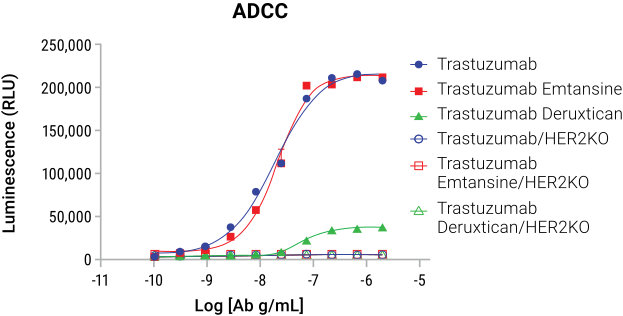
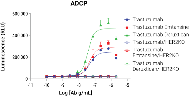
Fc Effector Activity Bioassays use genetically engineered effector cell lines to quantify ADCC and ADCP potency with precision, repeatability, accuracy and linearity to align with ICH guidelines.
ADCC involves immune cells targeting and destroying antibody-coated cells, while ADCP refers to the engulfing and digestion of target cells by phagocytes. Our Fc Effector Activity Bioassays are specifically designed to quantify the potency of these Fc effector functions, providing researchers with essential data to guide ADC development.
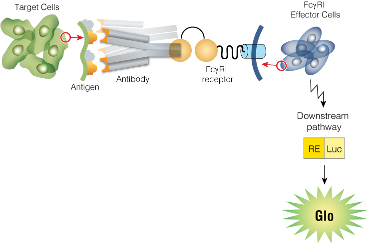
Measurement of ADC Fc Effector Activity using ADCC and ADCP Reporter Bioassays
Lumit® FcγR Binding Immunoassays
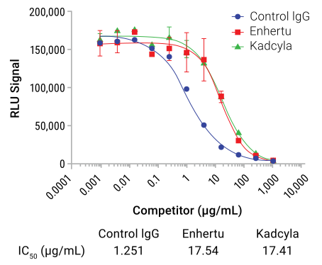
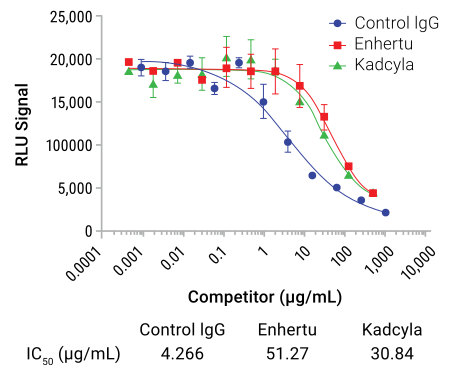
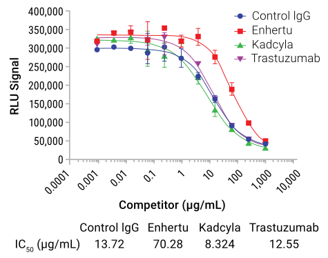
Lumit® FcγR Binding Immunoassays are your go-to ELISA alternatives. They are homogeneous, no-wash competition assays covering all major Fcγ receptors: FcγRI, FcγRIIIa, FcγRIIa, FcγRIIb, and FcRn. These receptors are key for influencing half-life, ADC pharmacokinetics, and immune activation.
Lumit® FcRn Assay
Lumit® FcγRIIa (H131) Assay
Lumit® FcγRIIIa (V158) Assay
Measure Complement-Dependent Cytotoxicity of Your ADC
Profile complement activation kinetics and predict therapeutic antibody efficacy with two complementary approaches:
- The Lumit® C1q Binding Assay quantifies the critical first step of C1q binding to antibody Fc regions. Get reliable results in under two hours with a simple, no-wash protocol.
- Then measure the downstream effect with the CytoTox-Glo™ Cytotoxicity Assay, which detects cell death resulting from complement-mediated damage.
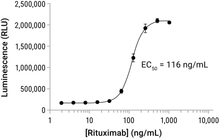
Daudi cells were treated with rituximab in RPMI 1640 + 12.5% human serum (the complement source). After 2hr, CytoTox-Glo™ was used to measure CDC on the Glomax® Discover.
Characterizing Fc effector function for your ADC? Explore the full bioassay portfolio.
How Do You Evaluate ADC Payload Specificity and Bystander Killing?
Analyzing the bystander killing effect, the impact on neighboring cells that do not express the target antigen, is essential for understanding therapeutic window and off-target risk in ADC development.
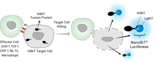
HiBiT Target Cell Killing Biossay measures death of a specific cell type within a mixed culture. Target cells express a HiBiT fusion protein that is released upon killing and binds extracellular LgBiT.
The Bystander Killing application uses three cell lines: wild-type HiBiT target cells (direct ADC cytotoxicity), wild-type "Dark" antigen-expressing driver cells (no HiBiT), and antigen-KO HiBiT cells (bystander killing). The ADC binds the "Dark" cell, internalizes the payload, and if it diffuses to kill neighboring antigen-KO HiBiT cells, the resulting luminescence is specific to bystander killing. Since HiBiT only signals upon membrane disruption, only dead antigen-KO cells contribute to the luminescent signal.
Poster: Novel Bioluminescent Tools for the Assessment of ADC Fc Function and Bystander Killing
What Custom ADC Research Services Does Promega Offer?
Promega's Tailored R&D Solutions team partners with ADC developers from lead optimization through IND-enabling studies, providing specialized analytical support powered by our bioluminescent assay platform.
Custom Assay Development
Tailor-made potency, internalization, and specificity assays designed for your ADC's unique target and payload.
Compound Screening
High-throughput identification and optimization of lead ADC candidates using validated cell-based readouts.
Mechanism of Action Studies
Detailed characterization of how your ADC kills target cells: direct payload effects, Fc effector function, and ICD.
Real-Time Cell Death Analysis
Continuous kinetic monitoring of ADC-induced apoptosis and necrosis over 72+ hours in live cell cultures.
Next-Gen Payload Analysis
Specialized assessment of degrader (PROTAC) and RNA-targeting ADC payloads with Promega's protein degradation platform.
Frequently Asked Questions
Antibody-drug conjugates (ADCs) are targeted cancer therapeutics built from three components: a monoclonal antibody, a linker, and a cytotoxic payload. The antibody binds a tumor-specific antigen, the linker attaches the drug stably for circulation, and the payload kills the cancer cell upon internalization. This architecture delivers potent cytotoxic effects directly to tumor cells while sparing healthy tissue.
ADCs engage the immune system through several mechanisms. The Fc region can engage immune effector cells by binding to Fc receptors on natural killer cells, macrophages, and other components of the immune system to destroy tumor cells. Certain payloads induce immunogenic cell death (ICD), releasing DAMPs and pro-inflammatory cytokines (IL-1β, IL-6, TNF-α, IFN-γ) that activate a broader antitumor immune response. These effects can be characterized using Fc effector bioassays, cytokine immunoassays, and ICD-specific readouts.
ADC payloads fall into three main categories: small molecule cytotoxins (the most common class, including microtubule inhibitors like maytansinoids and auristatins, and DNA-damaging agents like calicheamicins); protein degraders/PROTACs, which hijack the ubiquitin-proteasome system to eliminate target proteins; and oligonucleotides/RNA-targeting agents, an emerging class that silences or degrades specific intracellular RNA targets. Payload class determines mechanism of cell death, required drug-to-antibody ratio (DAR), and bystander killing potential.
Payload cytotoxicity is characterized using complementary cell-based assays on antigen-expressing target cells: cell viability assays (e.g., CellTiter-Glo® Assay) quantify overall survival via ATP; cytotoxicity assays (e.g., CellTox™ Green Assay) detect membrane integrity loss in real time; and apoptosis assays (e.g., Caspase-Glo® 3/7, RealTime-Glo™ Annexin V) distinguish apoptotic from necrotic death and capture killing kinetics. This multiparameter approach defines potency, mechanism, and therapeutic window.
ADCs bind target antigens expressed on cancer cells, triggering receptor-mediated internalization. Inside the cell, the acidic endolysosomal environment cleaves the linker and releases the cytotoxic payload, killing the cell from within. Some ADCs also engage immune effector mechanisms, including ADCC and ADCP, through their Fc region, adding an immunological component to their antitumor activity.
Two approaches are standard. pH-reactive dyes (e.g., pHAb Reactive Dyes) are conjugated directly to the antibody and fluoresce only in the acidic endolysosomal environment, providing a quantitative readout of internalization efficiency. Bioluminescent bioassays (e.g., the Antibody Internalization Bioassay) use a NanoLuc®-labeled anti-Fc Fab to detect internalized antibody-antigen complexes, with a membrane-impermeable inhibitor quenching any extracellular signal. Together, these tools support comparison of internalization across antibody variants and cell types.
Bystander killing occurs when a cytotoxic payload, released inside an antigen-positive cell, diffuses into the tumor microenvironment and kills neighboring antigen-negative cells. This is beneficial in antigenically heterogeneous tumors but raises off-target safety concerns. It is measured in mixed co-cultures of antigen-positive and antigen-negative cells using selective detection assays such as the HiBiT Target Cell Killing (TCK) Bioassay.